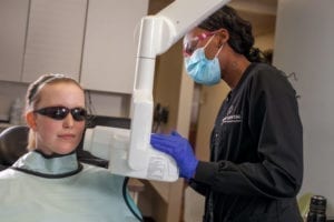
At Fuller Dental, we are dedicated to utilizing advanced technology and modern solutions to help our patients achieve a healthier, more functional smile. With advanced dental technology, we can enhance the patient experience and improve the treatment planning phase of your dental care.

X-rays are a focused beam of x-ray particles passed through bone which produce an image on special film, showing the structure through which it passed. This provides the familiar black, and white images of doctors and dentists use to diagnose problems and disease.
Without an x-ray of the whole tooth and supporting bone and gum tissues, there would be no way to detect infection or pathology that requires attention. In our office, we use digital radiography, which allows us to take x-rays using up to 90% less radiation than conventional film x-rays.
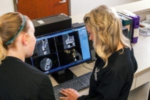
Using this technology, we are able to take an x-ray of your mouth by using a small sensor which records the image of your teeth and sends it to a computer. The result is a highly detailed image of your mouth that can easily be enhanced to better diagnosis dental concerns and determine the very best treatment for each case.
Digital Imaging Software

Fuller Dental uses digital imaging software in our office which allows us to take a digital picture of you and use our imaging system to predict how a particular treatment or cosmetic procedure would change the appearance of your teeth.
This software is beneficial for patients who are considering cosmetic procedures but are not sure if they’re ready for dramatic changes. Digital imaging also allows us to document your dental case and procedures very well. We take digital images of your face, teeth, and smile to provide us with a permanent dental record and to provide visual documentation of treatment.
CBCT Planmeca Imaging

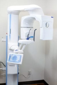
Cone beam imaging is a very valuable in-depth scan that captures many details that regular dental x-rays cannot capture. The CBCT imaging is typically used on patients that need an implant, orthodontics, and in some cases, when there are signs of a cracked tooth.
This scan allows providers to have a 360-degree angle of the tooth and the bone which surrounds the tooth. This 3D digital model of a patient’s anatomy is one of the fastest growing dental technology used in dentist offices and is becoming a standard piece in dental implant treatment planning.
